Home
Uncategories
Diagram Of The Muscles In The Forearm / Anatomy Forearm Anatomy Drawing Diagram : An upper arm muscle composed of 2 parts, a long head and a short head.
Diagram Of The Muscles In The Forearm / Anatomy Forearm Anatomy Drawing Diagram : An upper arm muscle composed of 2 parts, a long head and a short head.
Diagram Of The Muscles In The Forearm / Anatomy Forearm Anatomy Drawing Diagram : An upper arm muscle composed of 2 parts, a long head and a short head.. This muscle flexes the elbow and shoulder as well as supinates the forearm (i.e. Diagrams of the muscles of the forearm. From the arm muscle diagram above, the muscles of the arm that can be seen easily on the surface include biceps, triceps, brachioradialis, extensor carpi radialis longus, and deltoid. Most of these originate from the lateral epicondyle. The tendon then attaches to the most distal bone in the thumb.
For more anatomy content please follow us and visit our website: You can see in the arm muscle diagram above that there are important parts in arm muscles. Yoga anatomy anatomy study anatomy reference anatomy bones anatomy drawing hand therapy massage therapy physical therapy occupational therapy. It may last for a short time or even become a chronic problem. Once you're ready, you can try labeling the muscles for yourself using the blank forearm.
Easy Notes On Muscles Of The Anterior Or Front Of The Forearm Earth S Lab from www.earthslab.com Once you're ready, you can try labeling the muscles for yourself using the blank forearm. We think this is the most useful anatomy picture. Like the upper arm muscles, the forearm muscles can be divided into two parts: The large muscle of the upper arm is formally known as the biceps brachii muscle, and rests on top of the humerus bone. We'll go over the bones, joints, muscles, nerves, and blood vessels that make up the human arm. Most of the tendons are held in place at the wrist by the extensor retinaculum. Muscle general anatomy 12 photos of the muscle general anatomy general anatomy of a muscle, general anatomy of muscle fibers, general anatomy of skeletal muscle, muscle general anatomy, muscle general anatomy ppt, human muscles, general anatomy of a muscle, general anatomy of muscle fibers, general anatomy of. Muscles and tendons of the forearm pt 1
It is called lister's tubercle.
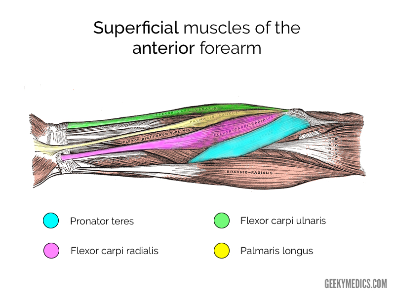
Like the upper arm muscles, the forearm muscles can be divided into two parts: As with the upper arm, the forearm is split into anterior and posterior compartment. Flexor carpi ulnaris, palmaris longus, flexor carpi radialis, and pronator teres. Diagrams of the muscles of the forearm. Most of these originate from the lateral epicondyle. The flexors, which lie on the inner side of the forearm and bend the wrist forward. Your arm muscles allow you to perform hundreds of everyday movements, from making a fist to bending your thumb. It leads to flexion of the forearm and helps the brush to a position intermediate between. The arm's curved shape comes from its major exterior muscles. Related posts of muscles of the arm and forearm diagram muscle general anatomy. You suspect a localized tearing of the origin of a muscle producing the equivalent of tennis elbow. the muscle most likely involved is the: To begin, spend some time looking at the forearm muscles diagram above. Related posts of forearm muscles diagram structure muscle relaxation anatomy.
Start studying muscles of the anterior forearm: The arms are the most used body parts and they can be subjected to much pressure and strain. We think this is the most useful anatomy picture. You can see in the arm muscle diagram above that there are important parts in arm muscles. Once you're ready, you can try labeling the muscles for yourself using the blank forearm.
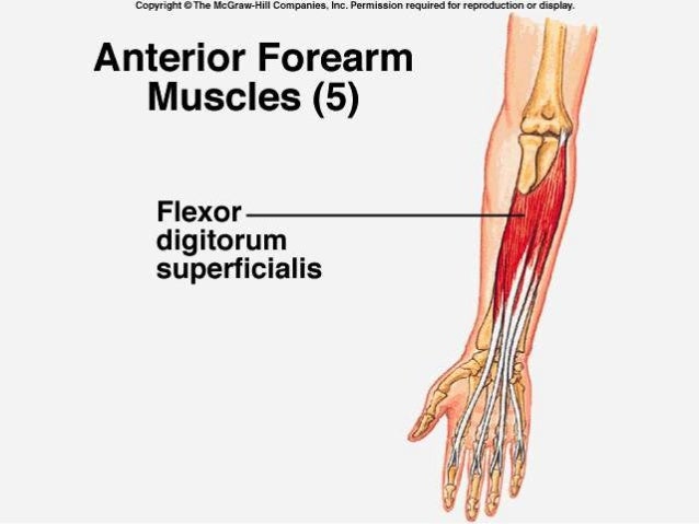
Muscles Of The Anterior Forearm Anatomy Geeky Medics from geekymedics.com There is no sensory loss in her forearm or hand. The photo on the left shows muscles that are deep to the ones on the right. Yoga anatomy anatomy study anatomy reference anatomy bones anatomy drawing hand therapy massage therapy physical therapy occupational therapy. It then travels around a prominent part of the radius bone that acts like a pulley. These muscles are all located on top of the forearm. The arm is one of the body's most complex and frequently used structures. Here you can see all the extensor forearm muscles clearly labeled. Its muscle belly is in the forearm and the tendon travels along the wrist and enters the third compartment of the band that holds the tendons in position at the wrist.
The arm is one of the body's most complex and frequently used structures.
An upper arm muscle composed of 2 parts, a long head and a short head. Muscle relaxation anatomy 12 photos of the muscle relaxation anatomy muscle relaxation anatomy, steps of muscle relaxation anatomy, human muscles, muscle relaxation anatomy, steps of muscle relaxation anatomy We'll go over the bones, joints, muscles, nerves, and blood vessels that make up the human arm. Wrist abduction is the lateral movement of your wrists to the right and left. Anatomynote.com found different types of muscles of arm diagram from plenty of anatomical pictures on the internet. While the primary function of your wrist extensors is wrist extension, these muscles also assist with the abduction and adduction movement of the wrist joint. Once you're ready, you can try labeling the muscles for yourself using the blank forearm. Yoga anatomy anatomy study anatomy reference anatomy bones anatomy drawing hand therapy massage therapy physical therapy occupational therapy. Learn with flashcards, games, and more — for free. To begin, spend some time looking at the forearm muscles diagram above. Learn vocabulary, terms, and more with flashcards, games, and other study tools. I've just switched over to a diagram to show you this muscle. Start studying muscles of the anterior forearm:
Superficial posterior muscles of the forearm posterior compartment muscles of the forearm. Pronator teres palmaris longus flexor carpi radialis flexor carpi ulnaris flexor digitorum. The photo on the left shows muscles that are deep to the ones on the right. Biceps are large muscle of the upper arm is formally known as the biceps brachii muscle, and rests on top of the humerus bone. Learn with flashcards, games, and more — for free.
Muscles Of The Forearm from image.slidesharecdn.com Overview diagram showing the labeled forearm extensor muscles forearm muscles (extensors) labeled and unlabeled. The arms are the most used body parts and they can be subjected to much pressure and strain. The long head originates just above the shoulder socket on the scapula and blends with the short head onto the radius bone of the forearm. As with the upper arm, the forearm is split into anterior and posterior compartment. Start studying muscles of the anterior forearm: This muscle flexes the elbow and shoulder as well as supinates the forearm (i.e. Once you're ready, you can try labeling the muscles for yourself using the blank forearm. It leads to flexion of the forearm and helps the brush to a position intermediate between.
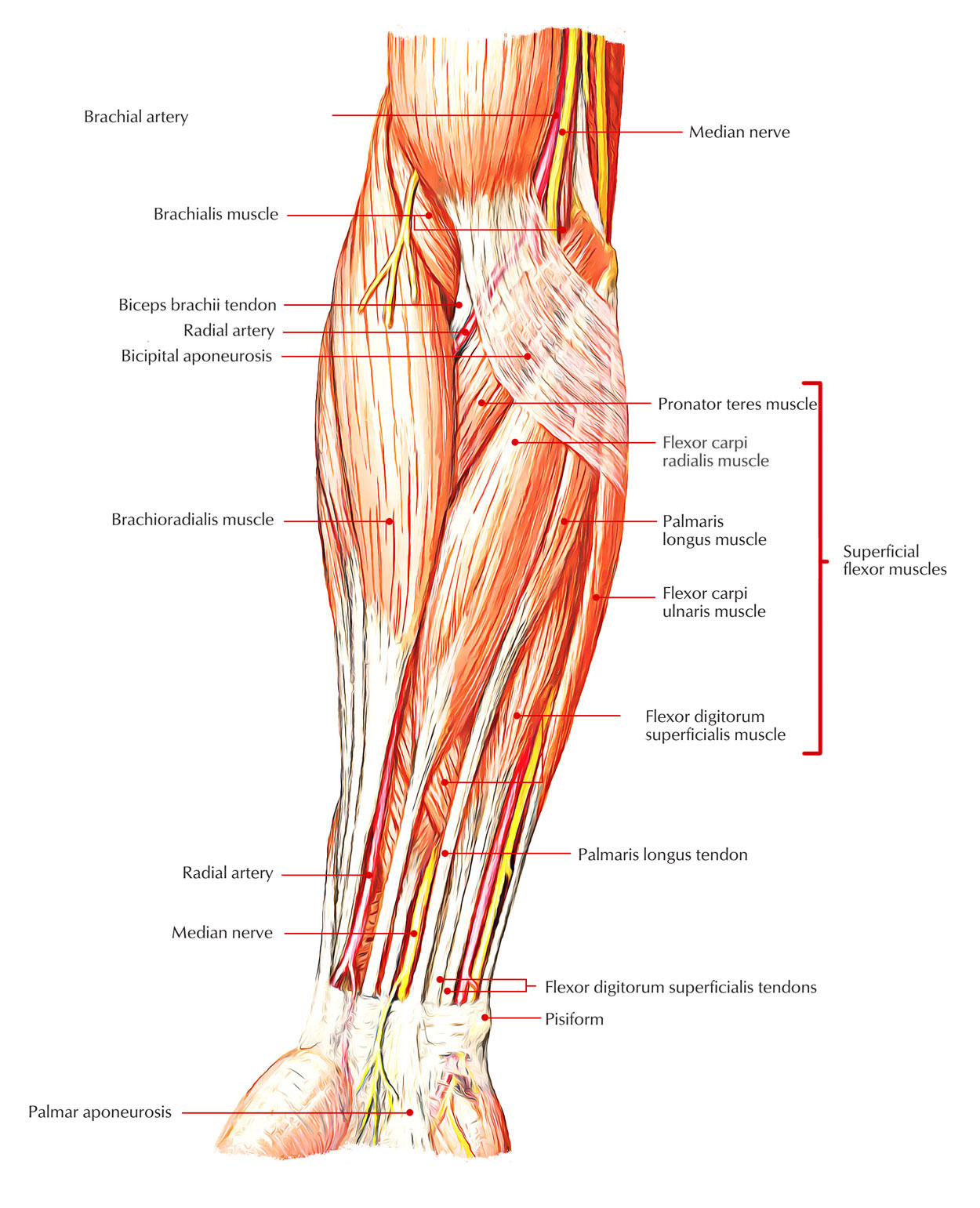
Forearm muscles in the anterior compartment are arranged in superficial, intermediate and deep categories.
Deep fascia of the forearm).—the antibrachial fascia continuous above with the brachial fascia, is a dense, membranous investment, which forms a general sheath for the muscles in this region; You can see in the arm muscle diagram above that there are important parts in arm muscles. This muscle flexes the elbow and shoulder as well as supinates the forearm (i.e. As seen in this forearm muscles diagram, the flexor muscles reside in the anterior compartment of the forearm, and are separated into the three following layers: Such pain can also originate from other parts of the body such as the neck or. There is no sensory loss in her forearm or hand. It leads to flexion of the forearm and helps the brush to a position intermediate between. The large muscle of the upper arm is formally known as the biceps brachii muscle, and rests on top of the humerus bone. You suspect a localized tearing of the origin of a muscle producing the equivalent of tennis elbow. the muscle most likely involved is the: Here you can see all the extensor forearm muscles clearly labeled. Pronator teres palmaris longus flexor carpi radialis flexor carpi ulnaris flexor digitorum. Learn vocabulary, terms, and more with flashcards, games, and other study tools. The flexors, which lie on the inner side of the forearm and bend the wrist forward.
0 Comments:
Posting Komentar